
Home
A comprehensive resource for safe and responsible laser use
US: Teen stares into laser pointer, has retinal damage
The unnamed teen initially had vision loss for several minutes due to flashblindness (looking into a bright light).
Five months later the boy went to an Ohio State University ophthalmologist due to continually-blurred vision with partial loss of vision in his right eye. Vision tests showed his left eye vision was normal. But if looking at text with his right eye, a single letter would be missing. When using only his left eye, or when using both eyes together, he could see the missing letter.
A standard clinical exam showed lesions in both eyes that were diagnosed as lesions in the macula, the area within the retina that we use for our central vision. The macula has the most and densest packing of light-detecting cones.
Tests done six months after the first doctor visit showed "marked improvement" in both eyes.
Further analysis was done with a custom-built adaptive optics optical coherence tomography scanning laser ophthalmoscope that is only one of five in the United States. This gives a very high-resolution view of the retina — much better than the human eye or more conventional retinal imaging techniques.
The AO-OCT-SLO image taken 11 months after the laser exposure showed damage to some of the macular cones. The ophthalmologist said "There's just nothing left there. The affected areas are devoid of cones."
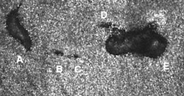
AO-OCT-SLO image of lesions A through E with small sites of cone loss (B, C, and D) in the teen's right eye. Each white dot is an individual cone cell, which is about 1/20th the width of a human hair. There are around six to seven million cones in the retina. Lesions A and E are about as wide as two hairs; lesions B-D are less than the width of a hair. Image source: Wolters Kluwer Health, Inc. / Ohio State University
Another AO-OCT-SLO image taken nine months later showed the lesions decreased in size, from 3.7% to 23.8% compared to the first image. "However, the longer-term prognosis is likely permanent scarring," according to the report.
Original report in Retinal Cases & Brief Reports, summarized in an Ohio State News story. Other stories about this appeared in Science Alert and Interesting Engineering,