
Home
A comprehensive resource for safe and responsible laser use
US: Teen stares into laser pointer, has retinal damage
The unnamed teen initially had vision loss for several minutes due to flashblindness (looking into a bright light).
Five months later the boy went to an Ohio State University ophthalmologist due to continually-blurred vision with partial loss of vision in his right eye. Vision tests showed his left eye vision was normal. But if looking at text with his right eye, a single letter would be missing. When using only his left eye, or when using both eyes together, he could see the missing letter.
A standard clinical exam showed lesions in both eyes that were diagnosed as lesions in the macula, the area within the retina that we use for our central vision. The macula has the most and densest packing of light-detecting cones.
Tests done six months after the first doctor visit showed "marked improvement" in both eyes.
Further analysis was done with a custom-built adaptive optics optical coherence tomography scanning laser ophthalmoscope that is only one of five in the United States. This gives a very high-resolution view of the retina — much better than the human eye or more conventional retinal imaging techniques.
The AO-OCT-SLO image taken 11 months after the laser exposure showed damage to some of the macular cones. The ophthalmologist said "There's just nothing left there. The affected areas are devoid of cones."
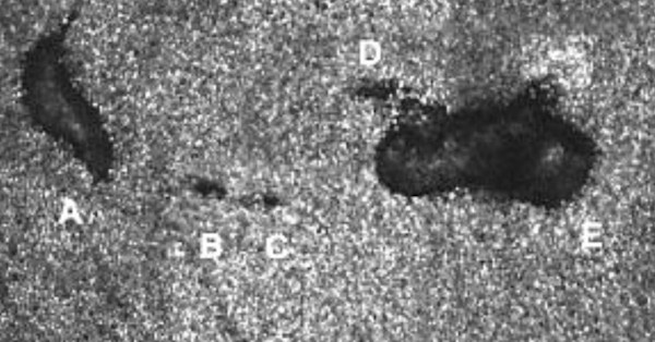
AO-OCT-SLO image of lesions A through E with small sites of cone loss (B, C, and D) in the teen's right eye. Each white dot is an individual cone cell, which is about 1/20th the width of a human hair. There are around six to seven million cones in the retina. Lesions A and E are about as wide as two hairs; lesions B-D are less than the width of a hair. Image source: Wolters Kluwer Health, Inc. / Ohio State University
Another AO-OCT-SLO image taken nine months later showed the lesions decreased in size, from 3.7% to 23.8% compared to the first image. "However, the longer-term prognosis is likely permanent scarring," according to the report.
Original report in Retinal Cases & Brief Reports, summarized in an Ohio State News story. Other stories about this appeared in Science Alert and Interesting Engineering,
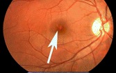
US: Rock concert fan injured by laser pointer-wielding crowd member
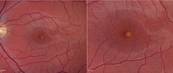
He was examined two days later at the Wills Eye Institute in Philadelphia. While slit-scan examination of the left eye showed no abnormalities, on an Amsler grid exam the patient drew a 2 mm circular spot. A fundus photograph showed a circular lesion in the fovea (magnified on the right):
Two weeks later the patient said there was some improvement in his vision. Fundus photography showed the lesion was smaller and less prominent; this was corroborated by optical coherence tomography (OCT).
The patient’s vision was expected to continue to improve over time.
From “Wills Eye Resident Case Series”, Jared D. Peterson, M.D. in the Review of Ophthalmology, November 7 2011. Introduction to case here; details of diagnosis, and discussion of eye injuries and treatment here.
Japan: Momentary exposure to Class 3B laser causes retinal injury
“In previously reported cases of retinal injury from red (He–Ne; 632 nm) laser pointers, the maximum output was 5 mW or lower (Class 3a), and gazing time was 10 s or longer. Higher-energy, green (532 nm) laser pointers are increasingly displacing red lasers and, here also, injuries have been reported. We report a case of retinal light damage caused, after a moment's gaze, by a high-output (Class 3b) green laser pointer unavailable to the general public in Japan that was brought from overseas.”
From “A case of retinal light damage by green laser pointer (Class 3b)”, Ueda, T., Kurihara, I. & Koide, R. Japanese Journal of Ophthalmology, July 2011, Vol. 55, Issue 4, pp 428-430, (2011) 55: 428. https://doi.org/10.1007/s10384-011-0031-5 First online July 1 2011.
Japan: Boy who routinely stared into a laser pointer develops lesion in one eye
At the age of 11, he had normal 20/20 (1.0) vision in the left eye, but 20/100 vision (0.20) in the right. Examination of the right eye showed a yellow lesion or fibrous tissue surrounded by a subretinal hemorrhage in the right macula. At age 13, examination showed the lesion was leaking on fluorescein angiography. At age 14, there was no change.
The doctors elected not to perform any treatment due to the patient’s age and mental condition.
From “Choroidal Neovascularization in a Child Following Laser Pointer-Induced Macular Injury”, Fujinami, K., Yokoi, T., Hiraoka, M. et al. Japanese Journal of Ophthalmology November 2010, Vol. 54, Issue 6, pp 631-633 (2010) 54: 631. https://doi.org/10.1007/s10384-010-0876-z First online January 21 2011.
US: Teen suffers central blind spot after older brother aims 50 mW laser at him
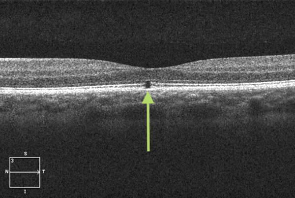
Tests at the Walter Reed National Military Medical Center Ophthalmology clinic one week after exposure showed no lesion visible to the eye (slit lamp exam) or with fundus photographs. However, Amsler grid tests indicated a central field visual defect in the left eye. Using more sensitive optical coherence tomography (OCT), a 56-micron disruption area was seen:
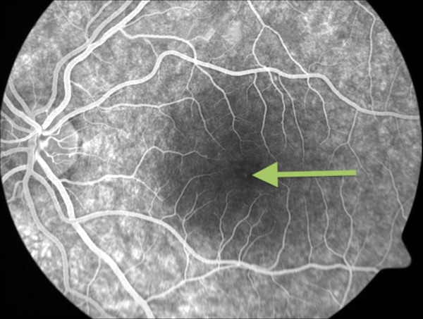
Intravenous fluorescein angiography displayed a barely detectable foveal window defect:
The diagnosis was that the blind spot was likely to remain, unchanged, and that treatment would not be necessary or effective,
Check-ups after two months and six months showed no change. However, after two years the teen no longer complained about a blind spot, and Amsler grid results were normal — despite OCT still showing the disruption area.
In an article describing the case, the authors concluded: “Our case represents a somewhat unique instance, where a moderate-powered [Class] 3B green laser produced visually significant retinal injury without correlating fundus findings on physical examination. The injury was only detectable by OCT and questionably fluorescein angiography…. Our case demonstrates the unpredictability of retinal findings in laser exposure in this power range, and the importance of OCT when evaluating patients who present with symptoms following dangerous laser exposures… If powerful lasers continue to be marketed as benign lights and their access to adolescent hands remains just a few keystrokes away, more ocular injury of this nature can be expected.”
From Military Medicine, Volume 180, Issue 3, 1 March 2015, Pages e378–e380, https://doi.org/10.7205/MILMED-D-14-00420
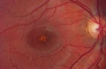
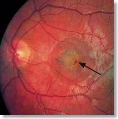
Greece: 9-year-old "repeatedly gazing" into laser causes hole in his eye
The most serious injury that the boy caused was a large hole in his macula, shown with the yellow arrows.
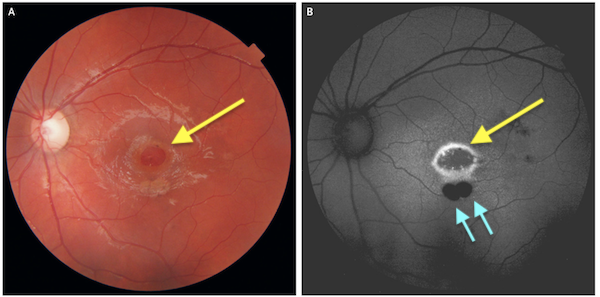
Two other areas of injury were not immediately visible in a funduscopic exam of the retina (photo A, using ordinary white light) but were clearly visible using fundus autofluorescence imaging (blue arrows in photo B, using a narrow wavelength of light). The round area to the left in both photos is the optic disc, a natural feature where the optic nerve begins — it is not laser damage.
The macula is where central vision occurs. The fact that the injury occurred in the macula indicates that the boy looked directly into the laser light with his left eye. Damage to the macula is serious as this area provides high resolution, color vision in the center of the visual field.
The injury reduced the boy’s vision to 20/100 in the injured left eye; his right eye remained at 20/20. The boy’s ophthalmologists felt the hole was too large and too much time had passed since the injury for surgery. (The doctors suspected that the boy had injured his eye at least a year earlier.) Because surgery might make things worse, causing a cataract without improving the macula, they “favored conservative management.”
There was no improvement in vision even 1 1/2 years after the injury was first presented to the ophthalmologists.
The power of the laser pointer, and other details of the incident, were not described in the one-paragraph report published June 21 2018. One of the authors told CNN the boy’s father “had bought the laser as a toy from a street merchant.”
From the New England Journal of Medicine (N Engl J Med 2018; 378:2420, DOI: 10.1056/NEJMicm1714488) Authors: Sofia Androudi, M.D., Ph.D., and Eleni Papageorgiou, M.D., Ph.D. Additional reporting by CNN. This story was picked up by many other news sites around the world.
US: Study examines four laser-caused eye injuries in children, at one medical practice
In a separate interview, one of the authors, ophthalmologist Dr. David Almeida, said these cases are “happening more frequently…. It was previously thought this was a one-in-a-million event. It's still probably a rare-to-uncommon reaction, but it's not a never reaction.”
All four children had foveal laser burns. Three of the children had potentially permanent vision loss. These are the cases:
- A 12-year-old boy looked into a green laser pointer for about a minute. He had decreased central vision in both eyes, with 20/20 vision in one eye and 20/30 in another. His vision and macular condition was found to be unchanged after 7 months.
- A 16-year-old teenager similarly had central vision loss in both eyes, after playing with a green laser pointer for about 30 seconds. He was first examined three days after the exposure, scars and atrophy were found on the retina. Two weeks later his vision has worsened. Visual acuity was 20/40 in both eyes with no improvement.
- A 9-year-old boy looked at the reflection of a green laser pointer in a mirror (essentially the same as a direct beam) for an unknown length of time. His vision was 20/50. He was treated with 1% prednisolone three times a day for two weeks. His vision improved to 20/30, but he still had “persistent abnormalities of the photoreceptors.”
- A 12-year-old boy looked into a red laser pointer for about 15 seconds. He had central vision loss, and 20/70 vision. He was given an injection of bevacizumab, which gradually improved his vision and symptoms. After 1 year, he had 20/20 vision.
The authors noted that laser pointers are more available, that users may not be aware of the dangers, and that some users may use pointers improperly.
Visible lasers less than 5 milliwatts (the U.S. legal standard for a laser to be marketed as a “pointer”) are considered to be generally safe due to the bright light reflex, which causes a person to blink and turn away from a bright light. So one question is why these children were injured by laser pointers.
One reason, according to the authors, is that “children increase their chance to retinal injury by staring at the laser beam without blinking or averting the eye for a prolonged duration.”
Another possible cause is that “the labeling of the power output of a laser point may be different from the device’s actual specifications.” They referred to a study of 122 laser pointers, where 90% of green pointers and 44% of red pointers were above the 5 milliwatt U.S. legal limit.
The study said that treatment options were “limited and also controversial.” Use of corticosteroids has shown “mixed results.” It may be enough to observe a patient over time, since many injuries will stabilize.
The authors recommended that laser pointer hazards “should be communicated to health professionals, school teachers, and guardians in an attempt to raise the public awareness of this emerging public health issue. Unsupervised use of these laser pointer devices among children should be discouraged, and there is a need for legislation to limit these devices in the pediatric population.”
From Retinal Injury Secondary to Laser Pointers in Pediatric Patients, Kunyong Xu, Eric K. Chin, Polly A. Quiram, John B. Davies, D. Wilkin Parke III and David R.P. Almeida, in Pediatrics; originally published online September 1, 2016; DOI: 10.1542/peds.2016-1188. A general interest article summarizing the study, with additional comments from Almeida and another ophthalmologist, is at HealthDay.com. The abstract of the Pediatrics article is below; click the “Read More…” link.
Click to read more...
Germany: Bus driver's eye injured by laser pointer aimed by child
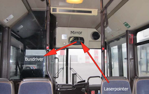
The 44-year-old driver stared into the laser several times, as he tried to identify the person holding the laser. He suffered blurred vision in his right eye immediately after the exposure, but waited 6 months before having his first complete eye exam.
The exam showed “spot-like retinal pigment epithelium disturbances temporal to the fovea of the right eye, with no abnormalities in his left eye.” The authors stated that “The subjective complaints and objective ophthalmological findings of this patient were consistent and strongly suggested that the repetitive exposure of the eye to the reflected laser spot 6 months previously had caused subtle but detectable injury to the macula.”
The authors concluded with two “Learning points”:
- “We suggest that no laser pointers of any class are made available to children, since they are unlikely to understand the risks of permanent retinal damage.”
- “For the safety of users and the general public, even low-energy handheld laser pointers should not be sold to children.”
The authors did not identify the location of the incident, but it may be Germany since three of the four authors’ institutions were in Germany. Additional analysis and commentary is below (click the “Read More…” link).
From Thanos S, Böhm MRR, Meyer zu Hörste M, et al. “Retinal damage induced by mirror-reflected light from a laser pointer” BMJ Case Reports. Retrieved online: 2015 Nov 05, doi:10.1136/bcr-2015- 210311.
Click to read more...
UK: Journal report of five children injured by laser pens
According to the abstract, “Clinically, three children had an acute vitelliform-like maculopathy which resolved to leave sub-foveal retinal pigment epithelium changes with reduced vision. One case was complicated by a choroidal neovascular membrane.”
- Case 1 was of a nine-year-old boy who on December 22 2013 was tested with normal vision of 6/5 (U.S. 20/17 -- better than 20/20) but on December 26 complained of vision loss and was found to have 6/12 (20/40) in the left eye and 6/15 (20/50) in the right eye. The family said he was given a laser pointer as a “toy” and had been playing with it on Christmas Day. The child denied looking directly into the laser beam. The family had three laser pens: a 57 mW blue 405nm, a 42 mW green 532 nm, and a 72 mW red 650nm. All exceeded the British Standard of 5 mW for a Class 3R laser. The boy was prescribed steroids. Nine months after the initial complaint, the best corrected vision was 6/9.5 (20/32), and optical coherence tomography showed persistent outer retinal layer disruption at the fovea. [The boy was later identified in press coverage as William Jackson, from Wadsley. Details are at The Star.]
- Case 2 was of an 11-year-old boy. He had decreased vision in both eyes of 6/7.5 (20/25). Eight weeks later he had sub-foveal retinal pigment epithelium changes. His vision was 6/12 (20/40) in the right eye and 6/15 (20/50) in the left eye. He said that a friend had aimed a laser into both of his eyes before the decreased vision occurred. The doctors were not able to examine what they characterized as the laser “toy”.
- Case 3 was of a 15-year-old girl. She aimed a laser pen into both eyes for 30 seconds. The next day she had scotomas (vision loss or spots) in both eyes. Her right eye was 6/7.5 (20/25) and her left eye was 6/6 (20/20). Upon examination, a vitelliform-like maculopathy (abnormality in the macula or central vision area) was seen. She did not return for follow-up visits.
- Case 4 was of an 8-year-old boy who had reduced vision of 6/12 (20/40) in his right eye, and normal vision of 6/6 (20/20) in his left eye. The right fovea was seen to have retinal pigment epithelial changes “consistent with laser burns.” The boy admitted he had played with a laser pointer a few months before, but said he did not point it directly at his eye.
- Case 5 was of a 13-year-old boy who had noticed declining vision in his right eye. It was found to be 6/36 (20/120); his left eye was 6/6 (20/20). He admitted aiming a laser pointer into his right eye. A fibrosed choroidal neovascular membrane was found at the right fovea.
The authors noted that “The retinal damage reported following such injuries is variable. This is due to variety of laser powers and wavelengths as well as ocular factors such as fundal pigmentation, blink responses, pupil size, and proximity of the laser burn to the fovea. Assessment of alleged laser eye injury requires accurate history and examination. Treatment for such laser retinal injuries is uncertain. Oral corticosteroids are sometimes administered.”
The authors stated that some laser devices are marketed as “toys”. They said they are aware of other children in the U.K. with retinal injuries from imported laser pointers. They conclude: “We suggest that children should not be given laser pointers as toys.”
From “‘Toy’ laser macular burns in children”, in Eye (2014) 1-4, by N. Raoof, TKJ Chan, NK Rogers, W Abdullah, I Haq, SP Kelly and FM Quhill. A downloadable PDF version is here. A story from the Bolton News gives some additional comments from author SP Kelly.
US: Child suffers eye injuries from adult misusing high-powered blue laser
The case was reported in JAMA Ophthalmalogy under the title “Ocular Safety of Recreational Lasers.” Authors Glenn Yiu, Sujit Itty and Cynthia Toth are with the Department of Ophthalmology at Duke University Medical Center in Durham, North Carolina.
They described the boy’s injuries as being caused by a Spyder III Pro Arctic “a class 4, high-powered 1250 mW laser that is manufactured from the 445 nm blue diode of a dismantled home theater projector and that is commercially available for online purchase from overseas.” This brand of laser is manufactured by the company Wicked Lasers; an 800 mW version was reviewed here.
In the case they described, “the adult directed the laser at the child’s eyes in jest, unaware of the harmful consequences.”
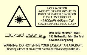
A copy of the safety label that appears on a Wicked Laser Spyder III Pro Arctic, containing the IEC and U.S. FDA-mandated wording for a Class 4 laser: “Avoid eye or skin exposure to direct or scattered radiation”
According to the authors, “imaging studies suggest that the laser damage was limited to superficial retinal vessels with no involvement of the underlying retinal pigment epithelium or choroid. To our knowledge, this is the first report of a continuous wave laser in the visible spectrum–damaging retinal vessels without affecting the retinal pigment epithelium, the site where damage from visible lasers typically occurs.”
They speculate that this may be caused by greater absorption of shorter wavelength lasers by hemoglobin, or a defocusing of the laser due to chromatic aberration and myopia in a child.
The authors conclude that “with the expanding use of lasers in nonoccupational or recreational settings, escalation of laser safety awareness and consumer laser regulations is paramount to prevent future ocular laser injuries.”
From JAMA Ophthalmology, published online January 09, 2014. doi:10.1001/jamaophthalmol.2013.5647. Thanks to Dr. David Hunter for bringing this to our attention.
Japan: Teen injured by LED pen "toy" held 40 seconds in his eye
A December 2006 incident has come to our attention. A 15-year-old Japanese boy suffered a retinal injury and visual loss after deliberately looking into a 5 mW violet (410 nm) light emitting diode for a total of about 40 seconds. The LED was in a pen was sold as a toy called “Secret Pen”. The toy appears to consist of an LED light which can excite ink that is invisible under ordinary light but which fluoresces under ultraviolet and near-UV light. The 410 nm wavelength caused photochemical damage to the retina.
According to a 2011 paper in Retinal Cases & Brief Reports, the LED was aimed into the teen’s eye from a distance of about 1 cm. It was held there for about 20 seconds as he deliberately stared into the light. This exposure was repeated the next day. About two weeks later, decreased vision (20/50 on the Snellen scale) was noted in the right eye.
Click to read more...
Germany: 11-year-old suffers eye injury from classmates playing
Afterwards, the boy could not see clearly and had a black spot in his visual field. He kept this from his parents for about three weeks, after which the boy was seen by Professor Stefan Dithmar and Dr. Stefanie Pollithy at the University of Heidelberg Department of Ophthalmology. Their diagnosis was “acute bilateral impaired vision and central scotoma.”
A journal article in Der Ophthalmologe has more information, but the full article requires a subscription. Jochen Pernstainer, who told LaserPointerSafety.com about the case, kindly provided several details from the article:
- The schoolyard exposure lasted several seconds
- The laser pointer was measured at 55 milliwatts
- The boy had impaired vision and a black spot on both eyes
- Nine weeks after the exposure his vision got a bit better
Fundoscopic photos of the 11-year-old boy’s left and right eyes. Larger versions can be seen here.
Dithmar told a local newspaper that the German Product Safety Act prohibits the sale of products that might cause harm to health, but “there is little that you cannot get on the Internet.”
Press report from die Rhein-Neckar-Zeitung (in German; an English Google-translated version is here). Journal article in Der Ophthalmologe, Vol. 109, No. 9 (2012), 907-910, entitled “Akute bilateral Visusminderung kit Zentralskotom bei einem 11-jährigen Jungen.” Thanks to Jochen Pernsteiner for bringing this to our attention.
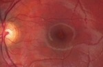
Switzerland: Boy injures self with 150 mW pointer
An examination two weeks later showed injuries to both retinas. There was severe vision loss in the left eye and 20/50 vision in the right. His left eye was injected with ranibizumab which helped improve vision to 20/25 after four weeks. The right eye improved on its own to 20/32.
The left eye clearly shows damage from a self-inflicted exposure to a 150 mW green laser pointer.
The report appeared in a letter published September 9 2010 in the New England Journal of Medicine.
UK doctors: Laser pointer damages youth's eyes
The burn site on the youth’s right eye